Research areas & fellows
DISCOVER’s ambition is to inspire and enable fellows to a research career bridging basic and clinical research. This will be done as a joint venture between BRIC and a number of clinical partner organizations.
You can read more about the fellows' PhD projects and aims in their individual project descriptions below.
DISCOVER fellows from first cohort:
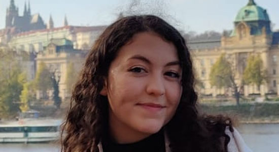
Name: Joana Castro
Nationality: Portuguese
Academic Background: MSc in Cellular and Molecular Biology and BSc in Biology, University of Porto
Project Title: Deciphering the molecular mechanisms of desmoplastic stromal reaction in solid tumours
Project Background: A Disintegrin And Metalloproteinase Domain 12 (ADAM12) is a member of the ADAM family, participating as both proteases and signaling mediators to control cell fate. This protein has been shown to be upregulated in a variety of solid tumours specifically in the cancer cells, in tumour tissue from breast cancer patients, and to be an indicator of disease stage. The mechanisms underlying ADAM12-mediated cancer progression are not fully understood. Previous results in our lab have shown a stimulatory effect of ADAM12 on both primary tumour growth and metastasis in breast cancer models in mice. Thus, we hypothesize that ADAM12, whether expressed on the cancer cell surface or released into the tumor microenvironment (TME), cleaves pro-tumorigenic molecules, such as the ECM remodelling protein Basigin, and mediates cancer-associated fibroblast (CAF) activation, thus increasing ECM deposition and aiding cancer progression. We hypothesize that the CAFs in the TME are activated by ADAM12 derived from cancer cells, resulting in excessive ECM deposition (desmoplasia) that can be addressed therapeutically to improve cancer therapy effectiveness.
Project Aim: In this study we aim to evaluate the therapeutic significance of ADAM12-associated ECM deposition in breast, pancreatic and colorectal cancer patients, as well as to unravel the molecular mechanism by which cancer cell-derived ADAM12 activates the tumor stroma and promotes ECM production. With the overall goal to identify new strategies to limit the desmoplastic response as a way to improve cancer treatments, we aim to investigate the clinical impact of ADAM12-associated ECM deposition in cancer patients, to reveal the causative link between ADAM12 expression and excessive ECM deposition, and to identify ADAM12-dependent paracrine mediators of CAF activation, using in vitro co-culture assays. The overarching objective is to find novel ways to inhibit the desmoplastic response and hence enhance cancer therapies.
Expected Outcome: The expected outcome of this project is to further consolidate the finding of a strong correlation between ADAM12 and desmoplasia. Moreover, we predict that the ADAM12-associated ECM content will exhibit a significant correlation to the clinical outcome of breast and/or colorectal cancer patients. Furthermore, we expect to reveal specific ADAM12-dependent changes in ECM expression, spatial distribution and/or structure, as well as changes in the CAF population and infiltration of various immune cells. Lastly, we hope to determine the mechanism whereby cancer cell-derived ADAM12 activates fibroblasts in the TME, including identification of one or more soluble factors that mediate the paracrine activation signal.
Contact: joana.castro@bric.ku.dk
Principal supervisor: Prof. Janine Erler

Name: Sanasadat Pourtabatabaei
Nationality: Iranian
Academic Background: Direct M.Sc. degree in Medical Biotechnology, University of Tehran
Project Title: Identifying and investigating the role of ribosome heterogeneity in pancreatic cancer
Project Background: The crucial complex of ribosomal RNAs (rRNA) and ribosomal proteins known as ribosome is responsible for translating genetic material into proteins. Ribosomes were thought to be housekeeping structures incapable of differentiating between various mRNAs. However, recent research from the Lund lab and others revealed that ribosomes have distinct compositions and modifications both at the protein and rRNA modification level, leading to the creation of ribosome subtypes (ribosome heterogeneity). The idea that ribosome subtypes offer an extra level of regulation to precisely control gene expression was strongly reinforced by subsequent research from the Lund group. Ribosomes' functional specialization is still being studied, therefore there is still much to learn about their functions in many biological circumstances, diseases, and the specific mechanisms that underlie them.
Project Aim: In this project, I will focus on translation in pancreatic cancer as one of the deadliest malignancies and a significant unresolved health issue. The main goal would be identifying ribosome heterogeneity and observing its effects on pancreatic ductal adenocarcinoma (PDAC) by focusing on alterations in rRNAs and ribosomal proteins. My hypothesis is that specialized ribosomes, with specific compositions and modifications at the protein and RNA levels, are positively chosen for translating mRNAs which promote pancreatic cancer. The project aims to characterize ribosome heterogeneity in pancreatic cancer. Specifically:
- How do ribosomes change in pancreas cells during pancreatitis and pancreatic ductal adenocarcinoma (PDAC) progression?
- Do different rRNA 2’-O-Me patterns and ribosomal protein heterogeneity exist, and how do they impact inflammation and tumorigenesis in the pancreas?
Expected Outcome: Based on the starting hypothesis, the expected observation is to detect significant effects of 2’-O-methylation of rRNAs and differential association of proteins to the core ribosome in pancreas tumorigenesis. These expected results would add to the knowledge of cancer- specific ribosomes in pancreatic cancer. This knowledge could be helpful for the development of new cancer prognostics and therapies.
Contact: sana.pourtabatabaei@bric.ku.dk
Principal supervisor: Prof. Anders Lund

Name: Phan Thu Han Le
Nationality: Vietnam
Academic Background: Pharmacist, M.Sc. of Pharmacy
Project Title: The role of N-WASP and its hotspot mutations in cancer
Project Background: N-WASP is a multi-domain molecule present both in the cytoplasm and in the nucleus which can promote actin polymerization, but probably also has actin-independent effects (Alekhina et al., 2017). Several published studies suggested a cancer-promoting role of N-WASP in intravasation, invasion, and metastasis (Gligorijevic et al., 2012; Hou et al., 2017). However, the loss of N-WASP in a mouse model for colon cancer led to complex responses (Morris et al., 2018). In mice with a keratinocyte-restricted knock-out of N-WASP, hardly any tumor formed and N-WASP deficient keratinocytes showed premature senescence in vitro. This effect correlated with the complex formation of nuclear N-WASP with the DNA methyltransferase DNMT1 and with the histone methyltransferases G9a and GLP, resulting in epigenetic suppression of the expression of the cell cycle inhibitor CDKN2A (Li et al., 2019; Li et al., 2018). These data indicated for the first time a cancer-relevant role for nuclear N-WASP.
Cancer is caused by copy number alterations and point mutations of driver genes resulting in cancer-promoting effects such as increased proliferation, impaired DNA repair, and evasion of anti-tumor immune surveillance. Based on earlier studies by our group and others and in silico analysis of more than 10 000 cancer genomes provided by the Cancer Genome Atlas (TCGA) we propose that hotspot mutations in N-WASP (gene name: WASL) have an oncogenic function in different malignancies
Project Aim: The aim of my project is to validate the cancer-promoting role of N-WASP hotspot mutations, to identify corresponding effector molecules, and to test the anticancer effect of an N-WASP inhibitor.
Expected Outcome: We expect to identify novel drug targets for personalized therapy of cancer patients carrying N-WASP hotspot mutations.
Contact: thu.lephan@bric.ku.dk
Principal supervisor: Prof. Cord Brakebusch

Name: Yen Thi Kim Nguyen
Nationality: Vietnamese
Academic Background: M.Sc. in Cancer Biology
Project Title: Phosphoproteome profiling for personalized leukemia therapy
Project Background: More than 75% of patients with Acute Myeloid Leukemia (AML) die within 5 years of diagnosis, primarily due to relapse and primary resistance toward current standard ChemoTherapy (CT) regimens including anti-metabolic and DNA damaging drugs. AML emerges through accumulation of genetic mutations including a few drivers that confer aberrant “oncogenic” activity of signaling and DNA Damage Response (DDR) pathways causally involved in malignant transformation and resistance toward CT. Consistently, clinical studies have demonstrated that cancer and AML patients, can be re-sensitized to CT by targeted inhibition of aberrantly activated signaling and DDR pathways. Although comprehensive genomics studies of large AML cohorts have identified novel therapeutic targets, these rarely identify the complete spectrum of druggable signaling and DDR molecules in AML patients, and have only led to approval of very few targeted therapies for AML treatment during the last decade. Hence, there is an unmet need to implement novel complementary diagnostic tools in the clinic to advance personalized therapeutic targeting of pathognomonic signaling and DDR molecules alone or in combination with conventional therapeutic regimens.
Project Aim: We aim to change the paradigm of explicit genomics-based AML diagnostics to an in-depth functional approach of combined genomics and Mass Spectrometry-based PhosphoProteomics (MS-PP) diagnostics. This novel approach is predicted to identify a broad spectrum of relevant druggable key signaling and DDR molecules that can be targeted by small molecule inhibitors to enhance CT efficacy, and ultimately improve clinical outcome of AML patients.
Expected Outcome: We will explore the potential of our existing MS-PP platform in a clinical setting for guided therapeutic targeting of oncogenic signaling and DNA damage response pathways in AML patients in combination with CT. This is expected to identify novel complementary drugs for personalized AML treatment, which can potentiate CT response, overcome CT resistance, and ultimately improve clinical outcome of AML patients as well as patients suffering from other cancer entities. Following the successful implementation and validation of the MS-PP platform, we plan to apply for additional funding to explore the full potential of our MS-PP platform to guide personalized combination therapies for relapsed/refractory AML patients in a future clinical trial at Rigshospitalet in collaboration with national and international AML centers.
Contact: yen.nguyen@bric.ku.dk
Principal supervisor: Clinical Assoc. Prof. Kim Theilgaard-Mönch

Name: Virág Gehl
Nationality: Hungarian
Academic Background: Bachelor of Science in Microbiology and Genetics (University of Vienna), Master of Science in Molecular Medicine (University of Vienna)
Project Title: Impact of IL-6 activation on tumour behaviour and therapeutic sensitivities in bile duct cancer
Project Background: Cholangiocarcinoma (CCA) is the second most common primary liver cancer arising from the hepatobiliary epithelia along the biliary tract. There is a tendency to increased incidence rates however, the reasons behind this are not clarified. Patients are often diagnosed at a late stage, as the tumour is already advanced or metastatic when surgical removal is not an option anymore. Palliative treatment is the standard-of-care for patients with unresectable tumours and some of the recurrent genomic alterations can be targeted specifically. However, resistance to chemotherapies and targeted therapies invariably arise.
There are different types of CCA tumours depending on their anatomical location; intrahepatic CCA (iCCA) and extrahepatic CCA (eCCA). iCCA is located inside the liver and it is associated with worse prognosis compared to the extrahepatic subtype. iCCA is driven by genetic and epigenetic alterations nevertheless, the liver provides a unique, often inflamed, milieu that further promotes the progression of the disease. In this dense desmoplastic tumor microenvironment the cytokine signalling networks are often deregulated and the interactions between the tumor and immune cells are altered. Elevated levels of the proinflammatory cytokine, interleukin-6 (IL-6) are associated with low overall survival, providing prognostic information. In addition, blocking IL-6 signalling increases the efficacy of chemotherapy in iCCA xenograft models.
Although, high inter- and intracellular IL-6 levels correspond to poor prognosis, it remains unresolved how exactly IL-6 activity impacts the biology of tumour cells, contributing to disease progression. I hypothesize that chronic IL-6 signalling results in transcriptional reprogramming in tumour cells, promotes resistance to chemotherapy but also leads to new vulnerabilities that can be therapeutically exploited.
Project Aim: Firstly, I will characterize the biological consequences of constitutive IL-6 signalling in iCCA by generating cell line models using CRISPR/Cas9. Then, I aim to identify targetable vulnerabilities acquired following IL-6 activation in iCCA by performing high-throughput drug screens. Lastly, I will investigate the impact of IL-6 signalling on tumour immunity and treatment response in iCCA by looking at samples from patients that underwent chemotherapy.
Expected Outcome: This project will provide novel insights into the underlying molecular mechanisms of IL-6-mediated tumour progression. The clinical relevance is ensured through applying primary patient-derived cell lines, tissue and blood samples and large clinical patient data sets to investigate the molecular and cellular consequences of active IL-6 signalling. This combined approach of experimental laboratory techniques and computational analyses will advance our understanding of tissue and systemic immunity in iCCA and help to identify new therapeutic approaches for iCCA.
Contact: virag.gehl@bric.ku.dk
Principal supervisor: Prof. Jesper Bøje Andersen

Name: Xian Xin
Nationality: Chinese
Academic Background: Bachelor of Science in Biological Science at Huazhong Agricultural University, China; Master of Science in Bioinformatics at Lund University, Sweden
Project Title: Investigation of mechanisms of epileptogenesis: integration of single cell omics from multiple animal models and human patients
Project Background: Neurodevelopmental brain disorders, such as schizophrenia (SZ), epilepsy (EP) and others, are one of the most frequent causes of patient disability and pose an enormous burden to the society. In most cases, the mechanisms how a neurodevelopmental disorder arises are unknown and furthermore current patient treatment procedures are very general affecting whole brain function and thus leading to a number of severe side effects. Substantial evidence shows that in many neurodevelopmental disorders certain types of neurons are more vulnerable than others, and that in a given brain disorder only particular neuronal subtypes exhibit signs of pathogenesis that might contribute to cognitive abnormalities. These data are supported by extensive research in animal disease models demonstrating that functional impairment of few neuronal subtypes can lead to disorganization of local neuronal circuits and underlie behavioral abnormalities, e.g. in SZ or developmental EP, including Khodosevich group’s own recent data showing that vulnerability of neuronal subtypes depends on the time of developmental insults.
In order to treat a disease, we need to understand what kind of cells become unhealthy, those cells that trigger the disease and how each cell type contributes to disease etiology and heterogeneity. Due to technology development last few years, all of these questions can be addressed by single cell omics approaches, including single cell transcriptomics, epigenomics, genomics and proteomics. Single cell technologies provide information in single (e.g. RNA) or multiple (e.g. RNA + open chromatin + protein) modalities for each cell in a complex tissue/organ. Thus, single cell technologies are powerful tools to study disease etiology and identify new targets for treatment and diagnostics.
Project Aim: The aim of this PhD project is to identify changes in gene expression and cell composition in cell-type- and region-specific transcriptomic and epigenetic data for mouse models and post-mortem human samples of SZ and EP, and to associate results with the physiological phenotypic data.
Expected Outcome: We will determine differences in single-cell transcriptome, open chromatin, and/or proteome data that are associated with neurodevelopmental disorders. These data will be used to identify those type(s) of cells and gene(s)/pathway(s) that contribute to neurodevelopmental disorders, which will finally be translated into new intervention strategies.
Contact: (email) xian.xin@bric.ku.dk
Principal supervisor: Prof. Konstantin Khodosevich
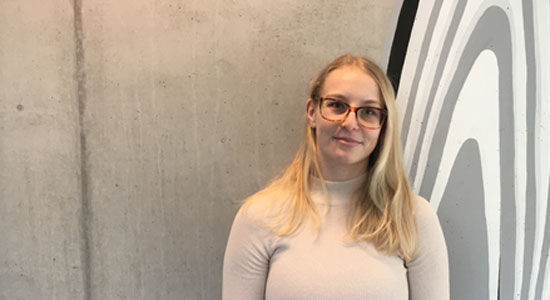
Name: Zala Zebec
Nationality: Slovenian
Academic Background: MSci in Neuroscience at University of Glasgow, with research placement year at the Institute of Clinical Neuroimmunology, LMU Klinikum, Munich.
Project Title: The role of IFNb-IFNAR signalling in Neuromyelitis Optica
Project Background: The focus of our group is neuroinflammation and investigating the role of traditionally immune genes in neuronal cells both under physiological conditions and in the context of neurodegenerative and autoimmune disease. Autoimmune disease is characterized by a failure of the immune system to recognize its own constituents as “self”, which allows an immune response against its own cells and tissues. Autoimmune diseases affect around 10% of the population and due to its chronic nature present a huge socioeconomic burden. Understanding the molecular mechanisms that lead to the mounting of an immune attack against “self”, is essential for the development of effective and safe therapeutics.
Neuromyelitis Optica (NMO) or Devic’s Syndrome, is a debilitating demyelinating disease of autoimmune origin. It is characterized by the presence of an autoantibody against a water channel – AQP4, expressed by the support cells of the brain – astrocytes. Without treatment, within 5 years, over 50% of the patients become blind and wheelchair bound, while 30% die. NMO shares many clinical features with multiple sclerosis (MS), however MS treatments have been shown to be ineffective or in some cases detrimental in NMO. Currently, NMO is mainly treated by general immunosuppressive drugs and plasma transfer. In contrast to patients with MS, IFNβ treatment is ineffective in NMO patients, yet the molecular basis of the distinct response in these two related autoimmune demyelinating diseases, remains to be fully elucidated. Improved understanding of molecular mechanisms that drive the autoimmune response against AQP4 is thus crucial for development of safer and more effective treatment options for NMO.
Project Aim: The overreaching aim of the project is to characterize the role of IFNβ-IFNAR signalling in molecular pathology of NMO by establishing novel disease models. Specifically, we aim to elucidate the molecular pathways that link IFNβ-IFNAR signalling and astrocyte homeostasis. Thereby, we hope to improve our understanding of NMO pathogenesis and identify new, highly specific therapeutic targets.
Expected Outcome: We expect to define the molecular pathways that links IFNβ-IFNAR signalling and AQP4 autoimmunity. Furthermore, we expect to elucidate how, why, and where the autoimmune response against AQP4 is mounted. Finally, we hope to translate our findings to humans, by examining whether our newly identified pathways are of relevance in patients and could therefore be used as novel therapeutic targets.
Contact: zala.zebec@bric.ku.dk
Principal supervisor: Prof. Shohreh Issazadeh-Navikas
DISCOVER fellows from second cohort:

Name: Ivona Cudina
Nationality: Croatian
Academic Background: M.D. (Integrated Studies of Medicine)
Project Title: The application of spatial technologies to the investigation of diseases with unknown pathogenesis
Project Background: Many human diseases are complex and involve the interaction of different cell types.
For example, in cancer the cancer cells can be divided in subpopulations with different properties based on different cancer driving mutations. Moreover, cancer cells interact with tumor stroma which consists of endothelial cells (blood vessels), innate and adaptive immune cells, fibroblasts, and extracellular matrix. Tumor stroma interactions were shown to affect cancer development and malignant progression and several stroma-directed cancer therapies have already reached the clinic.
Likewise, psychiatric disorders such as schizophrenia show complex impairment in neuronal networks and supporting cells, where coordinated impairment in multiple cell types underlies the etiology and symptoms. Schizophrenia is thought to be caused by a complex interplay of genetic and environmental risk factors influencing brain development. Recent research has instead concentrated on the molecular signatures of a more subtle pathology involving the functional state of specific cell populations and the architecture of cell-cell communication.
Therefore, analyzing cellular diversity in diseases will not only improve our understanding of disease pathogenesis, but also result in novel drugs development. In addition to the identification of different cell subsets in a tissue, also their location relative to each other is important. The shorter the distance between a ligand secreting cell and a cell expressing a corresponding receptor, the higher the chance of a functionally relevant interaction. Fields with overlapping influence of secreted factors can be recognized. Even single cells might be of highest disease relevance when placed at the right place, for example close to stem cells or a carcinoma in situ.
A similar network of interactions can be found in auto-immune diseases, where the interaction of lymphocytes and stromal cells is crucial for the development and responses of immune cells. Stromal cells play a role in inflammation, from induction to chronicity or resolution, via direct cell interactions and the secretion of pro-inflammatory and anti-inflammatory mediators. Vice versa, mediators released by immune cells during chronic inflammation cause changes in stromal cells, which might even develop a pathogenic phenotype. Understanding the significance and disease specificity of these interactions may lead to the development of new therapeutic options.
Project Aim: In this thesis project, I want to use and further develop latest spatial transcriptomics (ST) technologies and high content immunofluorescent imaging to investigate diseases, which are known to involve multiple different cell types and where the disease pathogenesis is unclear. By this approach I want to reveal different complex cellular and molecular pathways contributing to the development and progression of diseases. Such research could contribute to a better understanding of the pathogenesis, and to the identification of novel drug targets for therapy.
In collaboration with pathologists, bioinformaticians, and cell biologists I will analyze tissue sections from patients, describe their organization and develop novel hypotheses for the cellular and molecular mechanisms underlying the illnesses. To the extent possible, validation experiments will be carried out to confirm the hypothesis.
Expected Outcome:
The expected outcome of this research is to gain deeper insights into the intricate interactions between cells and receptors. By doing so, researchers aim to identify potential target points within disease pathways, enhancing our understanding of diseases' underlying mechanisms. This knowledge could lead to improved disease diagnosis and potentially more effective treatments. Additionally, the research seeks to uncover the spatial distribution of specific diseases, offering valuable insights into their localization within tissues and organs.
Contact: ivona.cudina@bric.ku.dk
Principal supervisor: Prof. Cord Brakebusch

Name: Justinas Daraskevicius
Nationality: Lithuanian
Academic Background: MD
Project Title: Targeting DNA replication and repair to overcome venetoclax resistance in acute myeloid leukemia.
Project Background: Acute myeloid leukemia (AML) is an aggressive hematological malignancy caused by genetic aberrations in hematopoietic stem and progenitor cells. Despite diagnostic advances and evolving molecular risk- stratified treatment strategies, intensive chemotherapy remains the cornerstone of curative AML treatment. However, most of AML patients are ineligible for intensive treatment due to advanced age or comorbidities leading to generally poor prognosis.
In 2018, FDA granted accelerated approval to BCL-2 inhibitor venetoclax (VEN) in combination with hypomethylating agents, such as azacitidine (AZA) for elderly/unfit patients with de novo AML, who are ineligible for intensive treatment. VEN-based regimens have become standard of care for these patients in USA and Europe. In addition, VEN/AZA-based regimens usually serve as salvage therapy for relapsed/refractory AML patients. However, significant number of responders eventually relapse due to acquired VEN resistance. Outcomes of AML patients who develop VEN resistance are dismal and discovering strategies to improve relapse-free survival is an unmet clinical need.
Genomic profiling of AML patients helps to understand the mutational landscape, but most of these patients do not have any targetable mutations and we lack effective biomarkers to identify individual patients, who likely will benefit from novel targeted agents. High-throughput ex vivo drug screening of AML cells to functionally identify drug response patterns represent a novel personalized medicine approach to improve AML patients’ outcomes, especially those who do not respond to standard therapy. It has been reported that ex vivo drug screening results can predict clinically meaningful drug responses and might be employed into clinical practice as surrogate biomarker of drug sensitivity. This approach could be used to identify novel synergistic drug combinations to overcome VEN/AZA resistance and combined with functional assays and -omics data provides us mechanistical insights about resistance mechanisms.
Project Aim: I aim to evaluate antileukemic potential, efficacy, and synergism of drugs targeting DNA repair and replication using high-throughput ex vivo drug screening platform and venetoclax-resistant AML patient samples. Functional studies will help to unravel mechanisms of venetoclax resistance and define the role of replication stress in VEN/AZA-resistant AML samples with the focus on PARP1/Wee1 and PI3K/AKT pathway inhibition. Finally, I aim to validate clinically relevant combinations using AML patient derived xenograft (PDX) animal models.
Expected Outcome: The most of AML patients eventually experience disease progression and become drug resistant. In this project, using AML patient samples together with the cutting-edge infrastructure I aim to identify and validate novel safe and effective treatment combinations that can overcome VEN/AZA resistance. Together, this approach will allow us to generate the information needed to establish next-generation AML therapies that hopefully will lead to improved survival of AML patients.
Contact: justinas.daraskevicius@bric.ku.dk
Principal supervisor: Prof. Krister Wennerberg

Name: Kartikey Saxena
Nationality: Indian
Academic Background:
Master of Science (M.Sc.) in Animal Biology and Biotechnology, University of Hyderabad, India
Bachelor of Science Honors (B.Sc. Hons.) Zoology, Sri Venkateswara College, University of Delhi, India
Project Title: Identification of novel targets in periphery of glioblastoma using spatial single-cell proteomics
Project Background:
Glioblastoma is the most frequent and malignant brain tumor, with 15,000 new cases per year in the E.U. and a median survival of approximately 14 months. Maximal surgical tumor resection is the first-line therapy, but in 90 % of the patients, glioblastomas recur in the resection margin within one year after surgery. Glioblastomas diffusely infiltrate the surrounding brain parenchyma, and complete resection is not possible. The cellular origin of recurrence is tumor cells left in the brain tissue – the real tumor periphery, escaping both surgical removal and intended removal by radiation and chemotherapy. In the tumor periphery, the diffusely infiltrating glioblastoma cells are found to be in intimate contact with tumor-associated microglia/macrophages (TAMs), reactive astrocytes, and neurons. These cells are capable of secreting cytokines, chemokines, and growth factors, thereby most likely having a pronounced influence on the local microenvironment, including the growth and migration of tumor cells. However, the potential proteins and pathways responsible for intercellular crosstalk in the tumor periphery are largely unknown due to limitations in obtaining tissue from this area and methodological limitations in identifying tumor cells in this area.
Project Aim:
The project aims to identify potential therapeutic target molecules in the tumor periphery using carefully collected patient biopsies and specific cellular biomarkers.
Expected Outcome:
This project will reveal large-scale spatial multicellular maps of glioblastoma, allowing the identification of potential therapeutic targets in the glioblastoma periphery.
Contact: kartikey.saxena@bric.ku.dk
Principal supervisor: Clinical Prof. Bjarne Kristensen
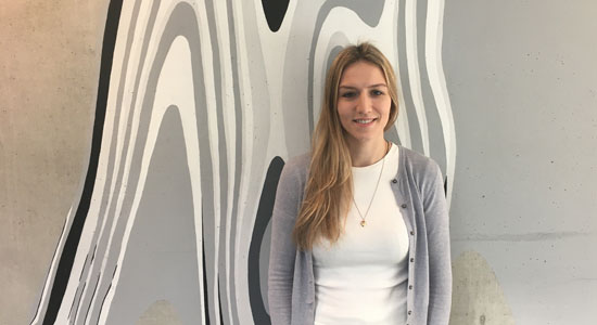
Name: Kyriaki Driva
Nationality: Greek
Academic Background: Bachelor of Science in Biology at Aristotle University of Thessaloniki, Master of Science at Karolinska Institutet in Stockholm.
Project Title: Lineage tracing-based discovery of treatments targeting drug resistant cell populations in ovarian cancer.
Project Background:
Among the gynecological cancers, ovarian cancer is the most lethal one in the western world. More than 70% of the ovarian cancer cases are diagnosed at advanced stage due to lack of screening strategies for early detection and less than half of diagnosed patients survive beyond five years. The standard model for classification of ovarian cancer is based on histopathology and grade, and approximately 90% of ovarian cancers are classified as epithelial. Epithelial ovarian cancer is a highly heterogeneous group of diseases rather than a single disease. At least 5 different histological subtypes of epithelial ovarian cancer can be identified, including high-grade serous, low-grade serous, endometrioid, clear cell, and mucinous as the most common ones, all associated with discrete pathology, origin, molecular profiles, genomic variability, and clinical response to treatments.
Among the epithelial ovarian cancer subtypes, high-grade serous carcinoma (HGSC) is the most diagnosed and lethal one. Standard care of HGSC combines debulking or cytoreductive surgery with platinum-based chemotherapy. However, despite the initial responsiveness of HGSC to platinum-based chemotherapy, it relapses very fast and becomes increasingly resistant. Underlying mechanisms of resistance to chemotherapy are yet to be fully understood. Intra- (within individual tumor) and intertumoral (within different tumors) heterogeneity within the same or different patients enhances the diversity and complexity of epithelial ovarian cancers along with the special and temporal heterogeneity, observed on molecular profiles of ovarian cancer tumors over time, possibly upon stress responses to specific therapeutic regimens. All this heterogeneity contributes to treatment failure and recurrence of disease. Thus, elucidating the role of heterogeneity in HGSC chemoresistance is crucial for designing therapies that can benefit the clinical outcome for HGSC patients in the future.
Project Aim:
The aim of this PhD project is to understand HGSC heterogeneity and unravel those cells that have the capacity to mediate drug resistance to standard chemotherapeutics. The key to achieving this goal is using appropriate tumor models. Thus, I am going to use patient-derived HGSC organoids because they recapitulate the genotypic and phenotypic features of original tumors. I am going to combine this model with cellular barcoding methodology which allows incorporation of unique barcode sequences that can be “read” both from the DNA and the mRNA of the cells. The barcoded organoid cultures will then be analyzed using different methods to identify differential lineage-dependent responses to standard chemotherapeutics and other various drug treatments.
Expected Outcome: By generating a multidimensional drug response profile for each lineage, I will then create novel drug combinations that can eradicate all the cells in the cell cultures. These drug combinations will be validated in vitro and functional assays will assist to unravel their mechanism of action. The 2 most promising drug combinations are going to be validated in vivo, using mouse models.
Contact: kyriaki.driva@bric.ku.dk
Principal supervisor: Prof. Krister Wennerberg

Name: Marvelmario Gabriel
Nationality: Indonesian
Academic Background:
Bachelor of Science in Biomedicine (Indonesia International Institute for Life Sciences, Indonesia). Master’s Double Degree: Master of Medical Science (Uppsala University, Sweden) and Master of Science in Molecular Medicine and Innovative Treatment (University of Groningen, the Netherlands), with research placement at the Karolinska Institute, Sweden.
Project Title: Tumor-macrophage dynamics and chemoresistance mechanisms in bile duct cancer.
Project Background:
Intrahepatic cholangiocarcinoma is a sinister bile duct malignancy. While it is classified as a rare cancer, most patients are diagnosed only at an advanced stage, with a very dismal prognosis of an average of 12 months survival. Worst, this survival time has not improved over the past decade. Most crucially, inter-patient benefit is highly heterogeneous.
For instance, in the clinic, we found that at diagnosis a cohort of patients with identical clinical characteristics and thus, expected to respond similarly to standard-of-care treatment. However, these patients diverged on chemotherapy into two extreme prognoses: one group surviving less than half of the median survival, whilst the other group surviving more than half of the median survival.
Preliminary analysis pinpointed when profiling the tumor cores that the differentially expressed genes between the two patient subgroups are predominantly derived from the tumor and myeloid cells, implying prominent cell-cell interactions between the two cellular compartments. Moreover, this is further supported by targeted transcriptomics, demonstrating strong dysregulations in crucial immune regulatory processes.
Project Aim:
The molecular and cellular differences in the tumor microenvironment are why the patients experience such extremely different prognoses. Hence, I aim to investigate various representative iCCA cell lines and their impact on monocyte/macrophage cell models, and vice versa through functional phenotypes and gene expression profiles to understand the cell-cell interaction (as seen in the patients). Next, I will perform ex vivo spatial transcriptomics analysis using patient samples. These data will be integrated with the in vitro findings.
Expected Outcome:
Through these approaches, we expect to unravel the crucial signaling proteins and mechanisms responsible in directing such extreme prognosis observed in clinics. I envision that this study will be key to understand the effectiveness of chemotherapy, and in the future may guide clinical decisions in choosing the best personalized approach for a patient (i.e., precision chemotherapy) and thereby, improving their prognoses and quality of life.
Contact: marvelmario.gabriel@bric.ku.dk,
Principal supervisor: Prof. Jesper Bøje Andersen

Name: Priya Khadgawat
Nationality: Indian
Academic Background: M.Sc. in Genetics and B.Sc. in Botany from University of Delhi
Project Title: A Novel Bispecific Antibody therapy for Acute Myeloid Leukemia
Project Background: Acute Myeloid Leukemia (AML) is an aggressive haematological cancer caused by genetic aberrations in haematopoietic stem and progenitor cells that generate immature AML blast cells in bone marrow (BM) leading to BM failure. Only 35-40% of younger/fit AML patients survive more than 5 years, due to relapse and primary resistance when treated with current standard chemotherapy regimens. Despite the advances in understanding the molecular mechanisms of AML development, only a limited number of novel effective drugs have been approved for AML treatment during the last decade. However, the vast majority of AML patients still fail to respond when treated with these novel drugs (i.e., FLT3, IDH1/2, BCL2 inhibitors, etc.) alone or in combination with conventional therapeutic AML regimens. Hence, there is an unmet need for novel complementary therapies such as immunotherapies that target AML effectively alone or in combination with conventional therapeutic regimens.
Currently, immunotherapies that target cancer cells specifically, such as ADCs (Antibody-Drug Conjugates) and bsAbs (bispecific recombinant antibody-based T cell engagers), have a huge therapeutic potential to improve the survival of AML patients. However, the development of bsAbs for AML treatment has been challenging due to the absence of AML-specific antigens. Hence, most current AML immunotherapies not only target AML effectively but also lead to substantial on-target toxicity on normal BM and blood cells, and to some extent non-hematological tissues. My Ph.D. project aims to develop novel bsAbs against specific targets to overcome therapy resistance and improve the clinical outcome of AML patients.
Project Aim: In this project, we aim to explore the therapeutic potential of bsAbs for AML treatment in a preclinical program including in vitro assays and established AML patient-derived xenograft mouse models. In collaboration, the laboratories of Prof. Ali Salanti and Kim Theilgaard-Mönch, recently identified glycosaminoglycan oncofetal chondroitin sulfate (ofCS) as a specific therapeutic target of AML cells not expressed by normal blood cells and non-hematological tissues. We aim to initially utilize this AML specific antigen based bsABs in an in-vitro setting, where we will define the therapeutic efficacy, toxicity profile, and T-cell dynamics of novel ofCS×CD3 bsAbs for subsequent validation in preclinical AML PDX trials. We will further follow this in-vivo by defining the therapeutic efficacy and toxicity profile of ofCS×CD3 bsAbs in pre-clinical PDX trials.
Expected Outcome: The expected outcome of this project is to develop a novel bispecific antibody based therapy for AML utilizing AML-specific antigen described above. Development and multi-level validation of this kind of therapy would potentially become a platform for a clinical program for AML patients and will further help in broadening the horizon of immunotherapies in AML and related malignancies.
Contact: priya.khadgawat@bric.ku.dk
Principal supervisor: Clinical Assoc. Prof. Kim Theilgaard-Mönch

Name: Qiuyue Wang
Nationality: Chinese
Academic Background: M.D., Master of Chinese Medicine (Dermatology) and Bachelor of Medicine
Project Title: Analysis of RhoA Driver Mutations in Cancer
Project Background:
Cancer is a widespread disease caused by mutations called driver gene mutations. These driver mutations are divided into oncogenic mutations, which are constitutively active versions of normal, often growth promoting genes, and mutations of tumor suppressors, which are tumor inhibiting genes that need to be deleted to progress cancer. Mutations observed in cancer, but not related to tumor formation or malignant progression, are called passenger mutations. Many oncogenic driver gene mutations have already been identified, resulting in targeted cancer therapies for some of them which complements classical treatment options. For even more mutations found in cancer, however, it is still unclear whether they are drivers or passengers. An example for this category of genetic alterations is RhoA E40Q, a mutation of the small GTPase RhoA found in about 1% of head and neck squamous cell carcinoma (HNSCC) patients.
In HNSCC patients, RhoA expression is often decreased. In the TCGA data set, more frequently than deep deletions of RhoA are E40Q mutations of RhoA. RhoA E40Q, a mutational hotspot in the RhoA gene and located in the Switch I domain of RhoA, which is crucial for the interaction with the effector molecule, thus providing a potential mechanism of action for a driver gene. Unpublished in vitro data of the Brakebusch group show now firstly that HNSCC with RhoA E40Q share a specific driver gene signature different from the majority of HNSCC, secondly that HNSCC cell lines expressing RhoA E40Q had higher RhoB levels and higher ROCK dependent phosphor- Myosin light chain (pMLC) amounts than corresponding cells expressing RhoA WT, and finally that the RhoA E40Q cells displayed increased migration dependent on ROCK. The reason for the higher RhoB levels in RhoA E40Q expressing HNSCC cell lines might be lower protein amounts of RhoA E40Q compared to RhoA WT expressing cells, which could be related to an impaired ability of RhoA E40Q to bind to effectors. Free, unbound RhoA is likely to be faster degraded than RhoA bound to effectors or regulators. These in silico and in vitro data strongly suggest that RhoA E40Q is promoting formation of invasive HNSCC by a RhoB/ROCK/pMLC/contraction pathway and can be rescued by ROCK inhibitor. Additionally, there are two other RhoA hotspot mutations RhoA T37S and G62E, which show a similar driver gene profile as RhoA E40Q, suggesting functional similarity.
Project Aim:
This project will establish the mice with a keratinocyte-restricted expression of RhoA E40Q, and induce the DMBA/TPA skin tumors in LSL RhoA E40Q RhoA fl/fl K5 cre. This study will Investigate the role of RhoA E40Q in skin cancer using a murine in vivo model and validate the effect of ROCK inhibitor in RhoA E40Q mice. In addition, this project will Investigate the driver gene function of RhoA T37S and G62E in HNSCC by testing in HNSCC cell lines with an induced KO of RhoA overexpressing T37S or G62E and investigate the effect of the RhoB/ROCK/pMLC/contraction pathway on the invasion of RhoAT37S and RhoAG62E in HNSCC, and to study the role of ROCK inhibitor in vitro.
Expected Outcome: This project will investigate the potential molecular mechanisms RhoA hotspot mutations play in tumorigenesis. We expect to reveal how RhoA E40Q promotes HNSCC formation in vivo and identify drug targets that specifically benefit patients carrying HNSCC with RhoA E40Q mutations.
Contact: qiuyue.wang@bric.ku.dk
Principal supervisor: Prof. Cord Brakebusch
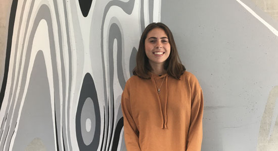
Name: Renata de Britto
Nationality: Brazilian
Academic Background: MSc in Biochemistry and BSc in Pharmacy
Project Title: How to handle VEXAS - a newly identified hematopoietic/inflammatory syndrome
Project Background: VEXAS (vacuoles, E1 enzyme, X-linked, autoinflammatory, somatic) is a new hematological and inflammatory syndrome. The disease has clinical features including hematologic abnormalities that could become a myelodysplastic syndrome (MDS) over time. Previous studies evaluating the bone marrows of patients with VEXAS noticed characteristic cytoplasmic vacuolization in myeloid and erythroid precursor cells. Despite the hematologic conditions, the patients also present systemic inflammation with relapsing polychondritis, giant-cell arteritis, polyarteritis nodosa or Sweet’s syndrome. VEXAS patients have mutations in the main E1 enzyme UBA1. The reaction catalyzed by E1 is an essential first step for all ubiquitin- dependent cellular process. Therefore, a deregulating in the activation step of ubiquitylation could impact the interaction between proteins. Most disease-defining mutations are limited to the pMet41 translation start codon for UBA1b. These results in the loss of the normally active cytoplasmic isoform UBA1b and leads to the appearance of a new enzymatically deficient isoform UBA1c. Meanwhile, the nuclear UBA1a isoform remains intact.
Project Aim: It is already known that VEXAS patients have decreased ubiquitylation, but how this process affects other proteins and its consequences are completely unexplored. As a result, more studies are required to evaluate the progression of the disease. Therefore, the main of this project is to understand the disease development of VEXAS and its impact on HSC self-renewal. To achieve this, we will 1) develop a model of VEXAS in mouse and human HSPC, 2) characterize the mutated HSPC in vitro and in vivo, and 3) identify positive and negative regulators of VEXAS with the aim to eradicate UBA1 mutated clones.
Expected Outcome: This project will not only yield novel biological insights into VEXAS but also identify targetable proteins that could be useful for the development of future treatment strategies aimed at targeting the aberrant HSC self-renewal properties of VEXAS mutated HSCs. Additionally, this project serves as a crucial bridge between clonal hematopoiesis and systemic inflammation, an area that demands further exploration and investigation.
Contact: renata@finsenlab.dk
Principal supervisor: Clinical Prof. Bo Porse
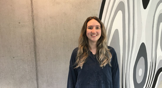
Name: Wibke Groenewald
Nationality: German
Academic Background: B.Sc. Biotechnology, M.Sc. Biotechnology with specialisation in Medical Biotechnology
Project Title: The role of dynamic rRNA 2’-O-methylation in brain development and disease
Project Background:
Tight control over cellular protein synthesis in space and time is critical for accurate stem cell differentiation. Providing the foundation for organ and tissue development, inaccuracies in protein quality or abundance during cell transitions can result in fatal developmental abnormalities or disease. Immaculate protein synthesis plays a critical role in the developing brain. The brain is the most complex tissue with a unique topical and functional geography. It consists of numerous types of neurons that require especially well-timed local cell fate decisions to reach the brains complexity without any mistakes.
The protein output can be regulated by genetic mutations, transcriptional or translational regulation and modifications. Effects of posttranscriptional, posttranslational and protein modifications have been extensively investigated, whereas the role of translation remains understudied. Translation of messenger RNA (mRNA) into protein is facilitated by ribosomes. Ribosomes are complex heterogenous machineries that, either alone or together with translation factors, enable regulation of translation. In recent years, the variety of ribosomal heterogeneity came into the focus of scientists, as evidence for their regulatory impact on translation is growing.
One impactful source of heterogeneity is 2’-O-methylation (2’-O-Me) of ribosomal RNA (rRNA). These methylations are installed by an enzymatic complex guided by specific small nucleolar RNA (snoRNA). The majority of rRNA nucleotides is methylated on all ribosomes, but a subset of methylations is variable between different cell lines, differentiation states and disease types, with potential regulatory function.
Recent studies from the Lund lab showed, that a single 2’-O-Me can change the translational profile of stem cells and alter specific signaling pathways thereby modulating cell fate decisions. Interestingly, three distinct methylation sites showed an increase during ectoderm differentiation which overlaps with observations in embryonic brain development in mice, but their role remains to be investigated.
Unsurprisingly, correct protein expression in all brain regions remains crucial throughout life. Abnormal protein accumulations and formation of fibrils in adult brain are main characteristics of diseases, such as in Alzheimer’s and Parkinson’s disease. The role of ribosomal heterogeneity on a 2’-O-Me level is unknown.
Project Aim:
My main hypothesis is, that dynamic ribosomal RNA modifications impact brain development by altering protein synthesis. In addition, I hypothesize that there are differences in specific 2’-O-Me in the ribosome pool of brain tissues with disease specific protein accumulations.
The project will pursue two main goals:
- Investigate potential effects of 2’-O-methylation of three specific rRNA nucleotides in human embryonic stem cells during ectoderm differentiation and in the development of brain organoids
- Unravel 2’-O-methylation patterns in brain disease and investigate potential site-specific methylation changes
Expected Outcome:
Exploring the role of changing 2’-O-Me in the human brains may benefit understanding its complex functions as well as the recognition and targeting of disorders. New insights into potential regulatory functions of single 2’-O-Me on protein synthesis and downstream effects in brain development will be gained. Therefore, a broad toolbox applicable for brain organoids, using histology and single cell technologies will be established. In addition, de novo mapping of 2’-O-Me patterns in brain diseases with abnormal protein accumulations may provide a unique perspective on the ribosome pool. Altogether, more information on the fine-tuning capacity of ribosomes by 2’-O-Me changes will result from this project.
Contact: wibke.groenewald@bric.ku.dk
Principal supervisor: Prof. Anders Lund
DISCOVER fellows from third cohort:
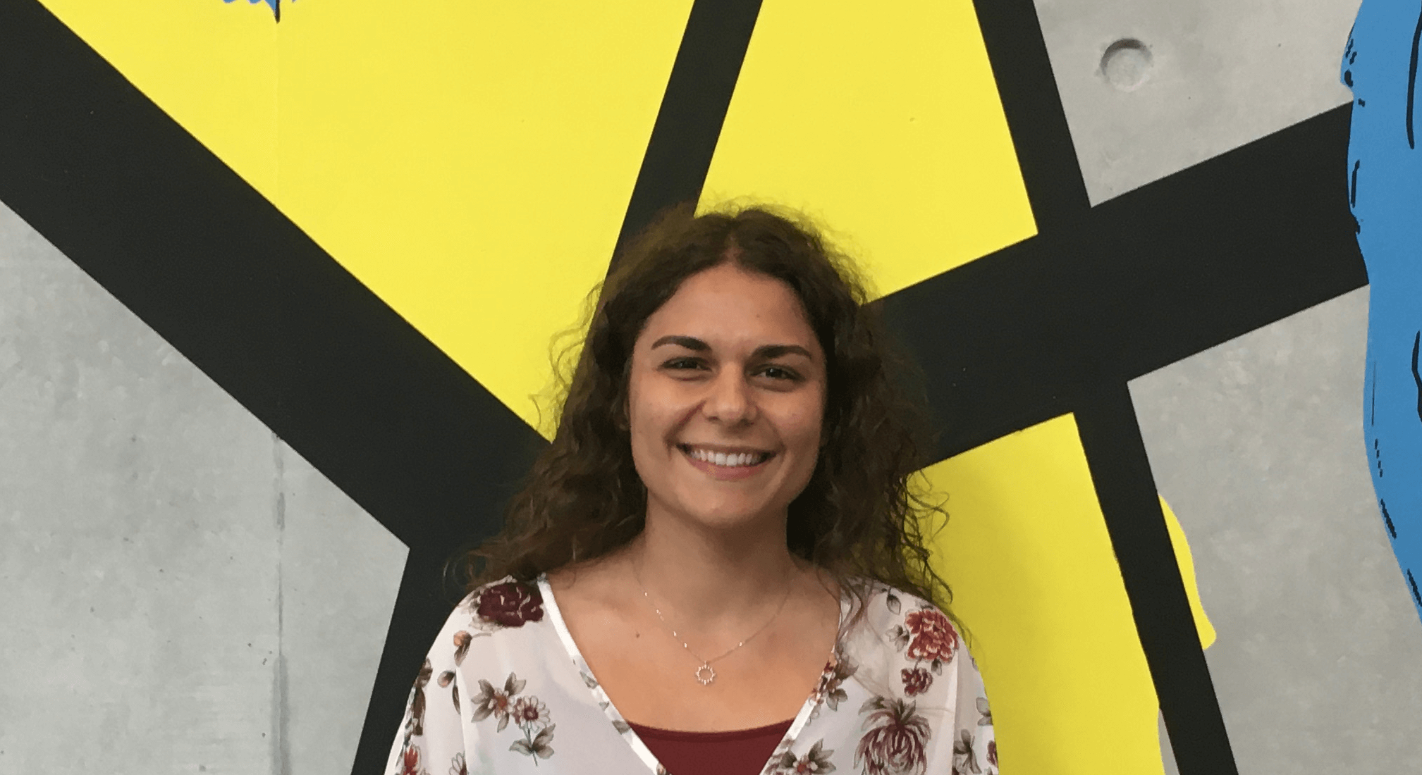
Name: Daniela Nunes
Nationality: Portuguese
Academic Background: MSc in Biomedicine at Karolinska Institutet, Sweden, and BSc in Cellular and Molecular Biology at NOVA School of Science and Technology, Portugal.
Project Title: Study the uPARAP receptor as a potential therapeutic target for immunotherapy in Acute Myeloid Leukemia
Project Background: Acute myeloid leukemia (AML) is characterized by the accumulation of immature poorly differentiated blast cells, resulting in the impairment of normal hematopoiesis. AML is responsible for most of the leukemia-associated deaths and although standard chemotherapy is initially effective, this treatment is extremely aggressive, and relapse is common. Therefore, more effective and less toxic targeted therapies are needed to increase long-term survival. One promising treatment is antibody-drug conjugates (ADCs) where an antibody is bound to a toxin released upon internalization, leading to the killing of the cancer cell.
Project Aim: Previous studies have identified the collagen receptor, uPARAP as being selectively overexpressed in AML and thus a good target for immunotherapy. Moreover, uPARAP is a recycling receptor highlighting its efficiency in taking up ligands. The main goal of this project is to assess the expression of uPARAP across the stages of the hematopoietic differentiation hierarchy in healthy cells and the context of AML. Furthermore, we will study the molecular role of uPARAP overexpression in AML and investigate whether we can specifically target these cancer cells with anti-uPARAP ADCs in vitro and in vivo.
Expected Outcome: Altogether, this study will gather information on the safety and efficacy of new anti-uPARAP ADCs and hopefully allow for their use as a less toxic treatment option for AML.
Contact: daniela.nunes@bric.ku.dk
Principal supervisor: Clinical Prof. Bo Porse

Name: Gaia Menichincheri
Nationality: Italian
Academic Background: B. Sc. Biological Sciences, M. Sc. Genetics and Molecular Biology
Project Title: Unravelling the RNA epitranscriptome governing genomic integrity and leukemogenesis
Project Background:
Gene expression regulation involves intricate mechanisms operating across multiple cellular mechanisms and pathways. Among these, evolutionarily conserved RNA chemical modifications are emerging as prominent regulators of central gene expression programs in health and disease. The epitranscriptome consists of over 170 different post-transcriptional RNA modifications, spanning all ribonucleotides. Importantly, RNA modifications significantly influence gene expression regulation, by affecting processes such as RNA stability, splicing and translation, with broad implications for development and disease (Roundtree et al., 2017). Advances in sequencing technologies have revealed how these dynamic modifications steer cellular fate. Pseudouridine (Ψ) was the first RNA modification discovered in 1951. Ψ is the most abundant chemical modification in RNA, accounting for 7–9% of the total uridine. Innovative work from our lab has highlighted a critical role of these Ψ synthases in regulating cells differentiation and stress responses, which is mediated by the deployment of small noncoding RNAs impacting translation
Project Aim:
Recent findings from our lab have identified PUS10, a Ψ synthase, as a key modulator of autoinflammation through dysregulation of transposable elements, remnant of viral infection driving evolution and genomic instability. Our findings indicate that PUS10 deficiency leads to increased global translation and activation of pathways such as DNA repair and G2-M checkpoint. This project aims to characterize the role of PUS10 in genome regulation through effects on coding and noncoding RNAs. Hematopoietic stem cells (HSCs) will be used as a model to study how PUS10-mediated Ψ impact genomic integrity through translatome reprogramming and other effects on DNA repair mechanisms. The project will further investigate the broader implications of Ψ dysregulation for altered HSC function in myelodysplastic syndrome (MDS), clonal hematopoietic disorders characterized by aberrant translation and genomic instability.
Expected Outcome:
This project will help us better understand the biological function of PUS10, particularly with regard to genome regulation. These insights may reveal novel therapeutic targets for age-related hematological disorders.
Contact: gaia.menichincheri@bric.ku.dk
Principal supervisor: Prof. Cristian Bellodi
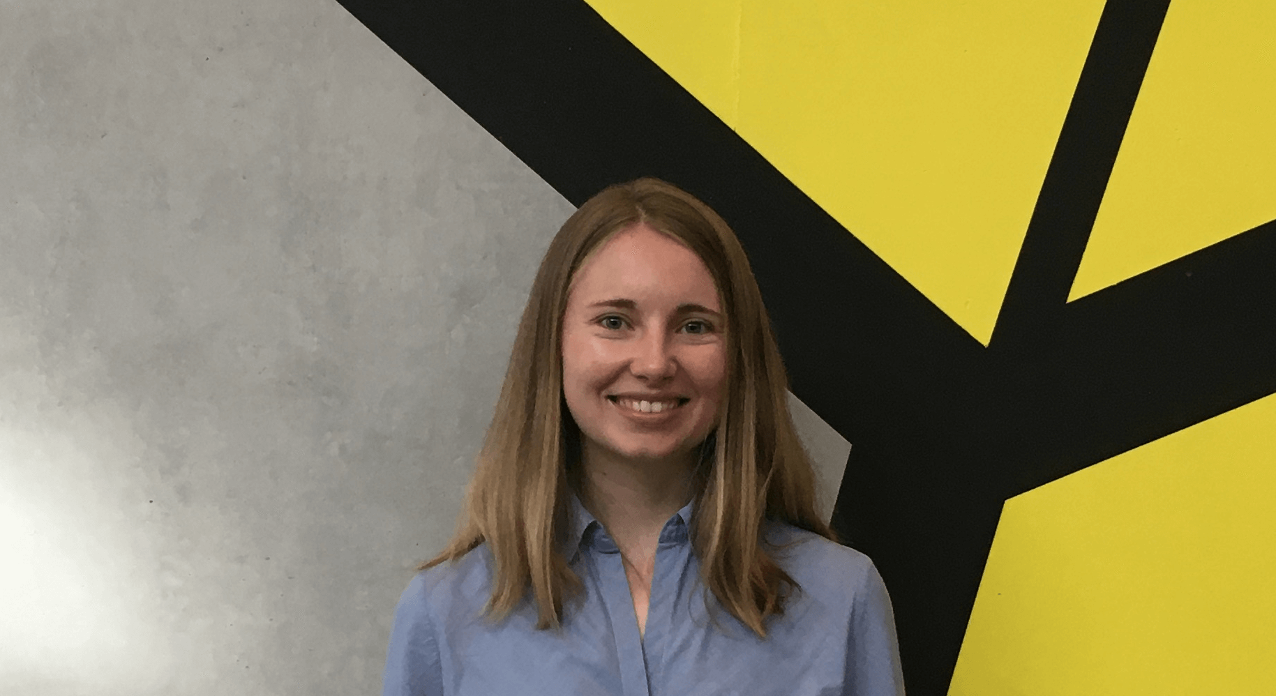
Name: Isabel Evi Naumann
Nationality: German
Academic Background:BSc. Molecular Medicine, M.Sc. Biomedicine
Project Title:“Transcriptional changes driving clinical heterogeneity in schizophrenia patients”
Project Background:
Schizophrenia is a severe psychiatric disorder affecting around 1% of the global population. Afflicted individuals suffer from a broad range of symptoms including delusions, hallucinations, social withdrawal, disorganized thinking, and cognitive deficits which all pose a severe burden on their health and socioeconomic situation. Despite the continuously growing knowledge about the clinical and molecular characteristics, risk factors, and disease-causing mechanisms, little progress has been made in developing novel, more effective treatments for schizophrenia. This is partly due to the immense complexity and heterogeneity of the disease in terms of its genetic and transcriptomic signatures, environmental risk factors, and clinical phenotype. Therefore, understanding the molecular mechanisms driving clinical heterogeneity is crucial to develop more personalized therapeutic approaches which would greatly improve the success rate of clinical trials and the efficiency of treatments.
Project Aim:
To unravel the molecular changes driving disease heterogeneity, we will apply cutting-edge single-nucleus RNA sequencing and spatial transcriptomic technologies to post-mortem brain tissue from over 300 schizophrenia and control patients from 5 different countries across the world. A recent single-nucleus RNA sequencing study published by the Khodosevich lab found large transcriptional heterogeneity and identified cortical upper-layer GABAergic interneurons as one of the major affected cell types. In my PhD, I will now delve deeper into the driving mechanisms and consequences of transcriptional heterogeneity in schizophrenia brains. The primary aim of this project is to link transcriptional changes to the clinical phenotype by integrating single-nucleus RNA sequencing data with the clinical metadata of these patients. Thereby, we hope to identify symptom-specific transcriptional changes and subgroup patients based on their molecular signatures. Symptom-specific alterations will then be further characterized in genetically modified mouse models regarding their functional and behavioral consequences.
Expected Outcome:
With our approach, we will be able to
- Identify common and shared transcriptional signatures and disease-associated compositional shifts across neuronal subtypes, layers, and individuals to better understand the disturbed neuronal functions and circuits in schizophrenia patients.
- Subcategorize patients based on specific positive, negative, and cognitive symptoms and characterize cell-type specific molecular signatures associated with each clinical subgroup. Thereby, we hope to identify novel potential biomarkers and therapeutic targets, paving the way to a more personalized treatment strategy that can greatly improve the lives of schizophrenia patients.
Contact: isabel.naumann@bric.ku.dk
Principal supervisor: Prof. Konstantin Khodosevich
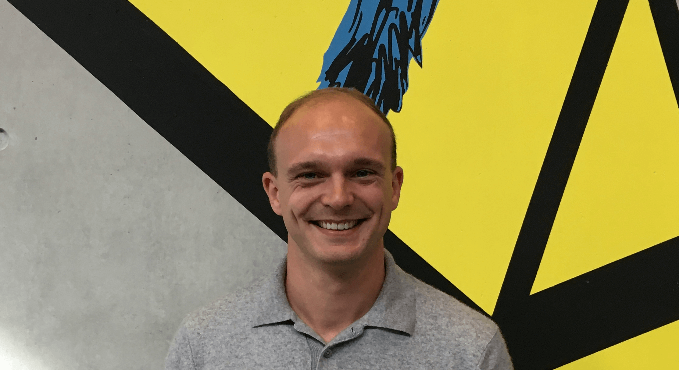
Name: Korbinian Schelzig
Nationality: German
Academic Background:
B.Sc. in Biosciences at University of Heidelberg
M.Sc. in Molecular Biosciences – Cancer Biology at the German Cancer Research Center & University of Heidelberg
Project Title:
Molecular insights into breakpoint formation in cancer patients: Structural variant analysis using Hi-C on biobanked clinical patient samples
Project Background:
DNA structural variations (SVs) affect more genes and are associated with worse outcomes than any other type of genetic alteration in cancer patients. SV formation is influenced by several mutational processes, including replication timing and chromatin folding, but their actual and relative contributions to disease initiation and progression remain unclear. Identifying SVs by whole-genome sequencing (WGS) from cancer patient samples depends on the availability of fresh tumor biopsies, however routine patient material is stored as formalin-fixed, paraffin-embedded (FFPE), making it inaccessible for WGS. Analysis of SVs from FFPE samples by immunohistochemistry or fluorescence in situ hybridization (FISH) is functional, but incomplete, as these are insensitive to small SVs, costly and labor-intensive. Recently, high-throughput chromosome-conformation-capture (Hi-C) as a method for detecting and quantifying spatially proximal DNA fragments from fresh-frozen samples, has been demonstrated as a tool to identify SVs with high sensitivity. This approach, when adjusted to the routinely stored FFPE samples, allows the exploration of SVs in a wide variety of cancer samples from many cancer patients and facilitates diagnosis and stratification of patients.
Project Aim:
To improve diagnostics of SVs in cancer patients, this project aims to a) optimize Hi-C on FFPE samples with a high sensitivity and assess the utility of FFPE Hi-C to investigate b) SV occurrence patterns and formation mechanisms and c) spatial localization of SVs from FFPE material.
Expected Outcome:
The approach will allow improved detection and investigation of SV formation and recurrence in large, clinically well-annotated and otherwise intractable biospecimens. Ultimately, this approach could allow deep investigation of SVs in larger biobanked patient cohorts and be added to already implemented diagnostics in the clinics and benefit cancer patients to receive more personalized treatment.
Contact: korbinian.schelzig@bric.ku.dk
Principal supervisor: Clinical Prof. Joachim Weischenfeldt

Name: Lisha Yuan
Nationality: China
Academic Background:
BSc. in Animal Science at Huazhong Agricultural University
MSc. in Basic Medicine at Sun Yat-sen University
Project Title: Single-cell Multiome in Decoding Human Hematopoiesis Along Development
Project Background:
Hematopoietic stem cells reside at the apex of the lineage hierarchy and can give rise to lifelong supplies of all blood lineages. The application of single-cell transcriptomic sequencing in the last decade has advanced our understanding that blood formation is a continuous process, coordinated by both intrinsic and extrinsic factors. Rather than being homogeneous, HSCs would be a heterogeneous population comprised of cells with existing lineage biases. However, it is largely unknown how early can lineage commitment be inferred in a primed state. Understanding the mechanism driving the phenotype of fetal HSCs, highly proliferative compared to their quiescent adult counterpart, will contribute to manipulating HSC expansion for transplantation therapy and enhancing the potency of HSC in healthier ageing.
Project Aim:
To explore this, we will leverage the recent Single-cell Multiome combined with cutting-edge computational methods to:
- Establish a comprehensive reference roadmap for human HSCs over a large window of time during development.
- Examine the dynamic regulatory patterns in priming HSCs commitment.
- Explore the temporal relationship between chromatin opening and gene expression to infer regulatory time lags along hematopoiesis.
Expected Outcome:
Through charting over 200,000 fetal hematopoietic cells from 50 fetuses aged from 7-18 weeks post-conception, this project will establish the first comprehensive transcriptional and epigenomic reference map of human developmental hematopoiesis and identify the impact of intricate interplay on the function of fetal HSCs. In the future, our study will extend to involve adult human bone marrow HSCs to systemically compare the discrepancies. This consortium of HSCs ontogeny established in this project could serve as a blueprint for harnessing the therapeutic potential of HSC and preventing the functional decline in HSC.
Contact: lisha.yuan@bric.ku.dk
Principal supervisor: Prof. Ana Cvejic
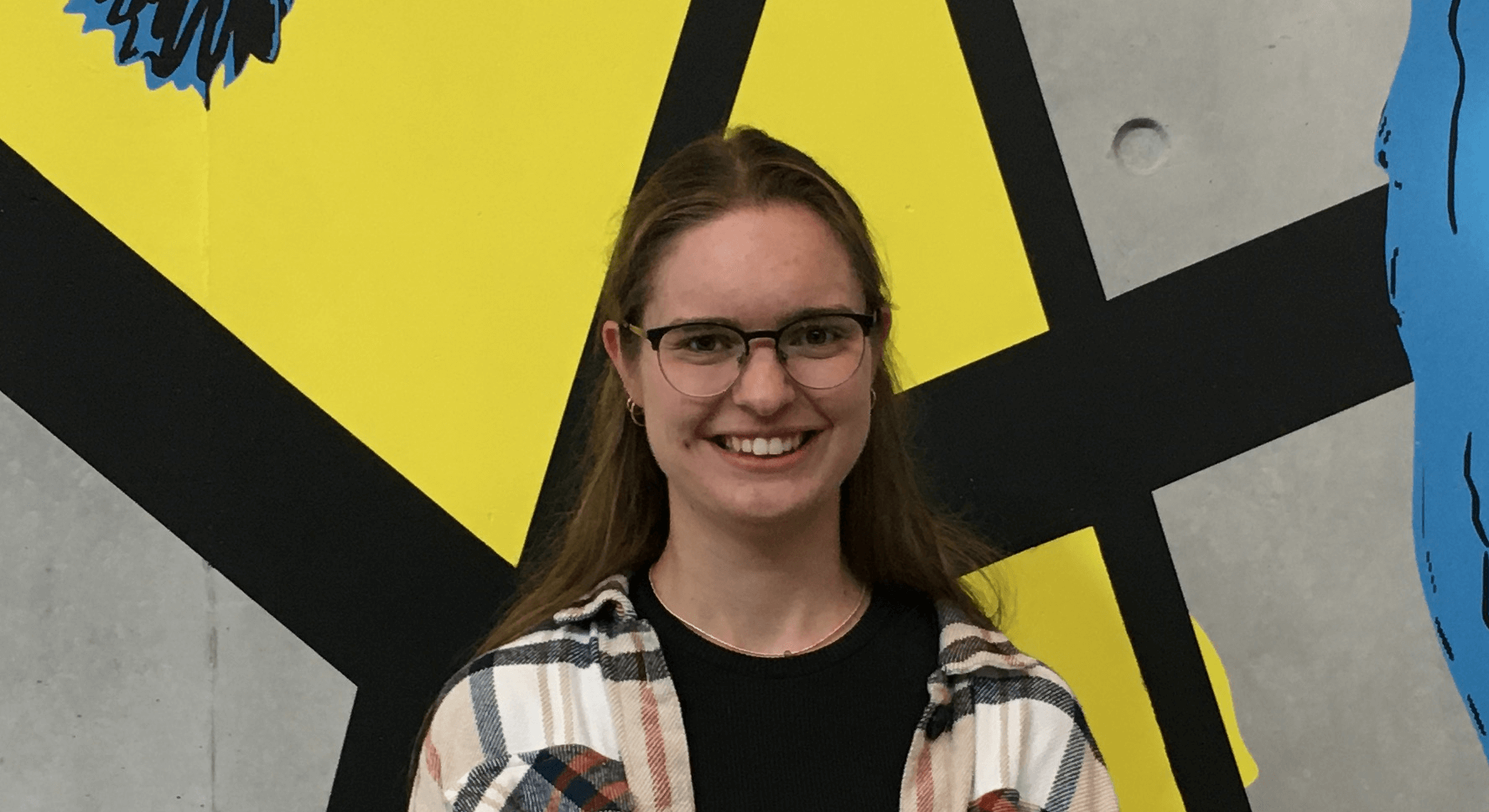
Name: Rosi Krebs
Nationality: German
Academic Background: BSc Biomolecular Engineering at Technical University of Darmstadt, Germany. MSc Biomedical Sciences (Specialization in Inflammation and Pathophysiology) at Maastricht University, Netherlands.
Project Title: Identifying and investigating the role of ribosome heterogeneity in glioblastoma
Project Background:
Dysregulation of translation is a characteristic of many cancers. The ribosome itself was long thought to have no intrinsic role in the regulation of translation. However, the accumulating evidence for ribosome heterogeneity, both at the level of ribosomal RNA (rRNA) and ribosomal proteins (RPs), suggests a regulatory role for the core ribosome itself, resulting in specialized ribosomes that affect the translation of specific messenger RNAs (mRNAs).
Modifications of the rRNA, such as site-specific 2'-O-methylations, contribute to ribosome heterogeneity. Changes in individual rRNA 2’-O-methylation levels have been observed in different tumor types and have been correlated with survival and prognosis. The Lund group and others have recently shown that individual rRNA modifications can contribute to the formation of specialized ribosomes. However, their specific role and the potential regulatory mechanism remain elusive.
Glioblastoma (GB) is the most common malignant brain tumor. Emerging data demonstrate altered translation in GB, but the focus in understanding translation in GB has thus far been on translation initiation, while translation elongation and potential intrinsic roles of the ribosome still remain unexplored.
Project Aim:
The objective of this project is to identify ribosome heterogeneity and its impact on translation and tumor progression in GB. Specifically, the project aims to:
- Identify ribosome heterogeneity and dysregulation of translation in GB.
- Investigate the role of relevant rRNA modifications and RPs in GB by using in vitro model systems.
- Investigate the mechanism by which individual 2’-O-me can affect regulation of translation.
To achieve this, patient-derived GB cell lines will be used and the ribosomes will be characterized regarding rRNA modifications and RP composition. In addition, small nucleolar RNAs (snoRNAs), that site-specifically guide rRNA modifications will be knocked out using a CRISPR library, allowing the identification of cancer-relevant snoRNAs.
Expected Outcome:
This project will provide novel insights into ribosome heterogeneity in GB and its role in tumor progression. In this context I expect to identify cancer-specific features of the ribosome, that could serve as interesting treatment targets and will be further investigated.
In general, this project will contribute to a better understanding of ribosomal heterogeneity and its consequences for specialized translation. The gained knowledge will not only be relevant for GB, but also for other types of cancer and the whole scientific field working on translational regulation and the ribosome.
Contact: rosi.krebs@bric.ku.dk
Principal supervisor: Prof. Anders Lund
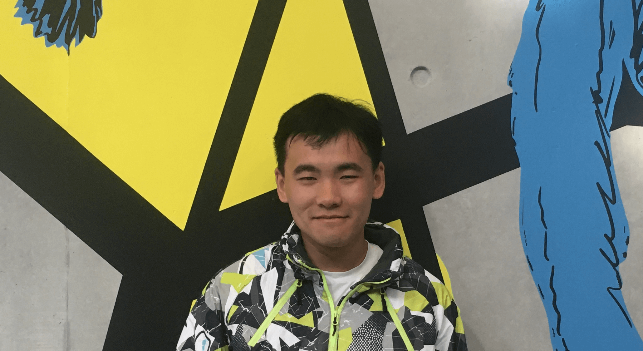
Name: Zixiang PAN
Nationality: China
Academic Background:
MSc. in Computer Science and technology (Computational Biology), Sun Yat-Sen University
BSc. in Telecommunication Engineering (Statistics Signal Processing), Hohai University
Project Title:
Exploring Hematopoietic Mechanisms in Healthy and Diseased Individuals through Single-Cell Mass Spectrometry
Project Background:
Reconstruct a single-cell proteomics (scp-MS) atlas of the hematopoietic system in both healthy and diseased mouse models to accurately define cellular states and differentiation trajectories.
Develop and optimize DIA-based scp-MS analytical pipelines, including:
- Quality control strategies for raw signal processing.
- Normalization methods tailored to non-amplified, intensity-based data.
- Denoising algorithms to enhance signal-to-noise ratio while preserving biological information.
Integrate scp-MS with single-cell transcriptomics (scRNA-seq) to:
- Investigate discordance between mRNA and protein expression levels.
- Refine classification of Lin⁻ stem and progenitor cell populations beyond conventional FACS gating strategies.
- Elucidate lineage-specific regulatory mechanisms in hematopoietic differentiation, including the role of protein-protein interactions in cell fate decisions.
Leverage artificial intelligence for computational tool development, aimed at:
- Enhancing data quality and interpretation in DIA-based scp-MS datasets.
- Building robust, cross-modal data integration frameworks for aligning unpaired scRNA-seq and scp-MS data.
- Addressing key challenges such as data sparsity, dynamic range differences, and low transcript-protein correlation.
Expected Outcome:
This project is expected to generate a comprehensive single-cell proteomic atlas of the murine hematopoietic system under both physiological and pathological conditions, providing unprecedented resolution in defining cellular states and differentiation hierarchies. The optimized data processing workflows for DIA-based scp-MS will establish a methodological foundation for future proteomics studies at single-cell resolution. Through multimodal integration of scp-MS and scRNA-seq data, we anticipate uncovering novel regulatory mechanisms underlying lineage commitment and the functional divergence between mRNA and protein expression. In addition, the development of AI-driven computational tools will offer scalable, generalizable solutions for noise reduction, normalization, and cross-modal data integration, paving the way for broader applications in systems biology. Ultimately, this work will not only deepen our understanding of hematopoietic regulation but also inform future diagnostic and therapeutic strategies for hematological disorders.
Contact: zixiang.pan@bric.ku.dk
Principal supervisor: Clinical Prof. Bo Porse
